Spine Imaging Course 2024
Stream this course now with the on-demand catch-up service
- An online spine imaging course featuring over a hundred practical MSK and Neuro cases
- Content will be delivered for consultant general radiologists andd senior STs with a practical and comprehensive update on advanced interpretation and reporting practice in spinal imaging
- Format: Short interactive presentations, followed by cases on PACS with discussion and Q&A
- CPD: 12 CPD credits in accordance with the CPD Scheme of the Royal College of Radiologists
Day One
60 minutes
Degenerative spine [including cord compression]
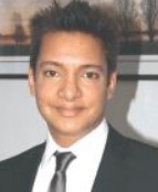
Dr Rajat Chowdhury
Consultant MSK Radiologist, Oxford University Hospitals NHS Foundation Trust
30 minutes
Paediatric spine

Dr Saira Haque
Consultant Paediatric Radiologist, King's College Hospital, London
50 minutes
Spinal canal abnormalities part 1: spinal cord
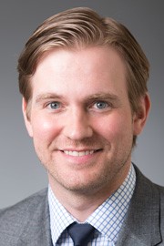
Dr Matthew Maeder
Assistant Professor of Radiology, Divisions of Abdominal Imaging and Neuroradiology, The Geisel School of Medicine at Dartmouth, Dartmouth-Hitchcock Medical Center, USA
50 minutes
Spinal Canal abnormalities part 2: Extramedullary and epidural

Dr Matthew Maeder
Assistant Professor of Radiology, Divisions of Abdominal Imaging and Neuroradiology, The Geisel School of Medicine at Dartmouth, Dartmouth-Hitchcock Medical Center, USA
50 minutes
Brachial plexus

Dr Sachit Shah
Consultant Neuroradiologist, National Hospital for Neurology and Neurosurgery, Queens Square, London
50 minutes
Thoracolumbar trauma
Dr Matthew Sarvesvaran
Consultant MSK Radiologist, Imperial College Healthcare NHS Trust
40 minutes
Spinal pathology in elite sports
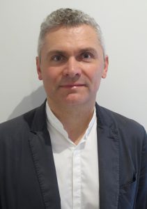
Dr Andrew Dunn
Consultant MSK Radiologist, Fortius Clinic
Day two
50 minutes
Spondyloarthritis and infection

Dr Ed Sellon
Consultant MSK Radiologist, Oxford University Hospitals NHS Foundation Trust
50 minutes
Surgical procedures and implants
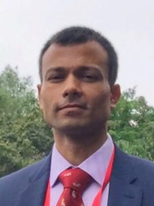
Mr Bedansh Chaudhary
Consultant Spinal Surgeon, Oxford University Hospitals NHS Foundation Trust
50 minutes
Non-vascular spinal intervention [including fusion imaging]

Dr David Wilson
Consultant MSK Radiologist, St Luke's Radiology, Oxford
50 minutes
Post-operative spine
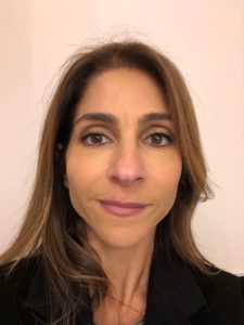
Dr Sarah Yanny
Consultant MSK Radiologist, Stoke Mandeville Hospital, Aylesbury
60 minutes
Cervical trauma [including the occipitocervical junction and anatomic variants]

Dr Qaiser Malik
Consultant MSK Radiologist, Basildon University Hospital
50 minutes
Spinal tumours [including marrow abnormalities]
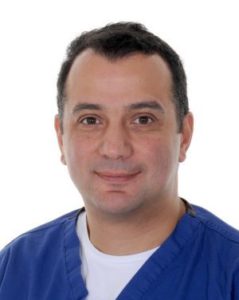
Dr Ramy Mansour
MSK Consultant Radiologist, The Ottawa Hospital, Associate Professor, University of Ottawa
The aim
To provide Consultant Radiologists with a practical, stimulating and comprehensive update on in depth interpretation and reporting practice in spinal imaging:
- practical (case-based learning);
- stimulating and challenging cases
- comprehensive (covering a full range of spinal imaging topics)
Course highlights
- Visiting lecturer from New Hampshire
- Faculty of Consultant Musculoskeletal Radiologists, Neuroradiologists and Spinal Surgeons who are experts in their fields and from different hospitals in the UK
- Interactive case-based approach to maximise learning, providing around 100 cases
- a greater appreciation – through collaborative discussion – of issues relating to spinal intervention
The content
Thirteen sessions over two days:
- Degenerative spine
- Spondyloarthritis and infection
- Post-operative spine
- Spinal canal abnormalities part 1
- Brachial plexus
- Thoracolumbar trauma
- Cervical trauma
- Spinal pathology in elite sports
- Surgical procedures and implants
- Non-vascular spinal intervention
- Spinal canal abnormalities part 2: extramedullary and epidural
- Paediatric spine
- Spinal tumours
Each session includes:
- A brief introduction by the session lead.
- Cases then reviewed by session lead whom will provide:
- Top tips
- Pitfalls
- Interactive questions
- Take home messages
Learning outcomes
By the end of the course, the delegate will have:
- a comprehensive understanding of spinal pathology in both specialist and district general settings;
- improved skills in interpreting emergency spine pathology;
- a more in-depth understanding of both routine and complex chronic spine abnormalities;
- learned tips and tricks to avoid common misses and pick up on subtle abnormalities, particularly in relation to topics that are often inconsistently reported;
- a greater appreciation – through collaborative discussion – of issues relating to spinal intervention
Course director

Dr Rajat Chowdhury
Rajat read Medicine at Oxford University and completed a Musculoskeletal Radiology Fellowship at Chelsea and Westminster Hospital, London and an Honorary Fellowship at the Nuffield Orthopaedic Centre, Oxford before being appointed Consultant at Oxford University Hospitals NHS Foundation Trust. Rajat has specialist expertise in all areas of diagnostic imaging and image-guided treatments relating to orthopaedics, spine, sports, rheumatology, sarcoma, bone infection, and trauma. Rajat has a particular interest in sporting injuries and works with elite athletes, professional footballers, cricketers, tennis players, and high-profile individuals.
Rajat is Radiology Lead for the postgraduate MSc. in Musculoskeletal Sciences at Oxford University and is an Honorary Senior Clinical Lecturer, supervising MSc. students. In addition, he sits on the admissions panel for Oxford University.
Rajat is Expert Advisor to the NICE Guidelines Committee for Osteoarthritis and was awarded a Lifetime Fellowship of the British Institute of Radiology. He was selected to represent the UK on the World Health Organisation Classification of Tumours Radiology Advisory Board. Rajat is author of several books including Radiology at a Glance and has contributed to the 4th edition of Gray’s Anatomy for Students. He is currently an Advisory Editor of the journal, Clinical Radiology and his current research interest is focussed on imaging and tissue pathology of soft tissue joint disease at Oxford University.
Faculty members

Mr B Roy Chaudhary
Mr B Roy Chaudhary is one the highest rated Neurosurgical Spinal Consultants in the UK*. He is a private Neurosurgeon/Spine Surgeon in Oxford having received gold medals, multiple scholarships and awards for his research and patient feedback on spinal and neurosurgical conditions. These include the Harry Morton Fellowship from the Royal College of Surgeons of England and the British Association of Spine Surgeons President's Fellowship Award. He is also a NHS consultant at Oxford University Hospitals having trained at Imperial College, London (M.Sc, Surgical Technology), University of Toronto (International Spinal Fellowship) and Cambridge, UK (NTN/Residency). *http://bit.ly/2fDh10M

Prof Elizabeth Dick
Prof Elizabeth Dick, MRCP, FRCR, MD, is a Consultant Radiologist and Honorary Senior Lecturer at Imperial College NHS Trust with expertise in Body MRI, Emergency, Trauma and MSK Radiology, subjects upon which she has published and lectured nationally and internationally.
She completed a two year Fellowship in Body MRI, during which time she earned her doctoral degree. She has published several books for Radiologists, and taught on numerous UK postgraduate courses to medical and paramedical specialists.
She is the Past President of the European and British Society of Emergency Radiology and was a Visiting Professor University of Sydney 2019-20

Dr Andrew Dunn
Dr Andrew Dunn completed radiology training on the Mersey School of Radiology in 2005, and completed a fellowship in musculoskeletal imaging from the University of Toronto in 2006. Andrew is a consultant and honorary clinical lecturer at the Royal Liverpool University Hospital.
Dr. Dunn has a specialist interesting in the imaging of sports injury, and provides imaging services to many Premier League and Championship football clubs, Super League Rugby Clubs and other elite sports institutions throughout the Northwest region, and was a member of the MSK imaging team for the London 2012 Olympic Games.
Andrew maintains an active role in education, teaching on the Mersey School of Radiology and the Northern Deanery Fellowship of Sports and Exercise Medicine training programme.

Dr Ramy Mansour
Dr Mansour the Clinical Lead of MSK Imaging at Oxford University Hospitals. He is the Royal College of Radiologists (RCR) Global Ambassador and the former RCR BSSR Travelling Professor 2019-2021. Dr Mansour was the MSK team Lead of the European Diploma in Radiology (EDiR) from 2019 to 2023. He trained at Oxford University Hospitals and obtained the Royal College of Radiologist Fellowship (FRCR) in 2005. He is an invited speaker in national and international conferences, and he delivered more than 400 lectures in countries in Africa, Asia, Europe and the Americas. He published articles and abstracts in the medical literature and regularly asked to peer review articles for national and international journals. He is a member of the British, European and Asian Musculoskeletal Societies and member of the Skeletal Society of Radiology (SSR) and International Skeletal Society (ISS). He is an active member of the ISS Outreach Committee and the regional coordinator for North Africa and the Middle East. He is a member of the RCR International Committee and the ESSR Educational Committee. He is the winner of the RCR inaugural International Travelling Fellowship Award for 2017-19.

Dr Matthew Maeder
Dr. Matthew Maeder is an Assistant Professor of Radiology at the Dartmouth-Hitchcock Medical Center. He completed medical school at the University of Cincinnati, followed by Radiology residency training at the Lenox Hill Hospital in New York City. He is dual fellowship-trained in both Body MRI and Neuroradiology, both at Dartmouth. His interests include diseases of the central nervous system, head and neck, as well as the liver and prostate. Dr. Maeder is experienced in a variety of procedures including CT-guided biopsies of the lungs, liver, and kidneys as well as spinal procedures including vertebroplasty, sacroplasty, facet joint synovial cyst rupture, and spine biopsy. He is currently on the faculty at Dartmouth specializing in Abdominal Imaging and Neuroradiology. He is the Director of the Body MRI fellowship.

Dr Qaiser Malik
Dr Malik is the lead for MSK imaging at Mid and South Essex NHS Foundation Trust.
He is a Honorary Senior Lecturer for University College London Medical School. He has served on the Scientific Programme Committee for 7 years for the Royal College of Radiologists and was the Newsletter editor for 2 years.
He has also served on the British Society of Skeletal Radiologists Executive Committee responsible for Education and Research including awarding of research funds and professorships. More recently he is an active member of the UKIO conference committee organising the MSK stream of the conference in 2020 and 2021.

Dr Sachit Shah
Dr Sachit Shah is a consultant in diagnostic neuroradiology at the National Hospital for Neurology and Neurosurgery at Queen Square. Dr Shah studied medicine at University College London, and undertook postgraduate training in medicine at Hammersmith and Guy’s & St Thomas’ Hospitals, and the National Hospital for Neurology and Neurosurgery. Following this, he trained in radiology at Imperial College Hospitals, before undertaking subspecialist training in neuroradiology on the Pan-London Neuroradiology Fellowship. In addition to wide ranging expertise including stroke, pituitary and dementia imaging, Dr Shah has a specialist interest in the imaging of peripheral nerve and muscle disorders. Dr Shah also holds an academic post at the UCL Institute of Neurology, and is a lecturer on the UCL Advanced Neuroimaging MSc programme.

Dr David Wilson
Dr David Wilson trained at King’s College Hospital, London and the Oxford Radcliffe Hospitals. His primary interest is in the application of modern imaging techniques to disorders of the locomotor system and spine intervention. He has undertaken original work in the application of diagnostic ultrasound to joint, muscle, and soft tissue disease with particular attention to joint effusion and congenital dysplasia of the hip. He has over 20 years of experience in vertebroplasty and is the author of publications on multicentre controlled trials on the treatment of insufficiency fractures. He has established innovative training courses in the UK in musculoskeletal ultrasound in Oxford and Bath. He teaches internationally and is a leader in the development of ultrasound in musculoskeletal disease and injection techniques in the spine.

Dr Sarah Yanny
Sarah studied Medicine at the Royal Free and University College Hospital, during which time she undertook a BSc degree in infectious disease, before attaining membership of the Royal College of Physicians in 2006. She trained in Radiology at the Norwich Radiology Academy, after which time she completed a Fellowship in Musculoskeletal Imaging at the Nuffield Orthopaedic Hospital, Oxford.
She was appointed to Buckinghamshire Healthcare NHS Trust in 2011 where she leads the MSK service and is Clinical Governance co-lead.
She has an interest in education and research, teaching on several regional courses and has published several musculoskeletal papers in peer-review journals as well as reviews and book chapters.
Delegate comments from 2024 Course
- Excellent content and breadth of coverage
- Very good sessions with scrollable cases and very well organized
- The cases and the lecture deliveries all very good.
- It was online, interesting cases to scroll by myself, excellent speakers – all good to me!
- The sessions on tumour/spa/brachial plexus were very good for me
- Great speakers covering very good content with good PACS support
- Very good content and delivery by most lecturers
- I hope to implement all the good tips that I’ve got from the lectures and systematise my approach in reading spine and brachial plexus MRI.
- The topics will contribute to improve my performance and be more confidence in reporting acute spines
- This course updated my knowledge and improved my understanding
Delegate comments from previous courses

Access to cases for our imaging events
Our imaging courses are very much an interactive experience. Presentations are kept to the minimum and then you'll be into the fully featured cloud based DICOM viewer, looking at cases, feeding back your findings using our interactive tools. You'll get immediate feedback and learning points from our expert faculty member.
- Attendance of the course includes access to the database of cases associated to this event on our server at PostDICOM.
- Full access to each case with a full toolset to open, view and manipulate each case alongside the faculty but on your own screen!
- You will maintain your access to the resource throughout your 60 day catch-service period too.
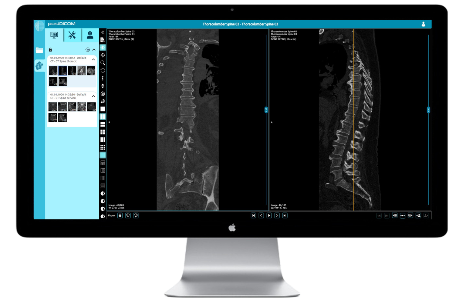

Sample the DICOM viewer here. A window will load below the buttons (best demonstrated on a computer rather than mobile device)
When will I receive my course login?
We will contact you by email one week before the course takes place with all the necessary links and joining information.
We will re-send the links the day before the course.
If you have not received an email from us please contact us at webinars@infomedltd.co.uk and we will respond ASAP.
Will I need any special software to partake?
NO. Infomed shall provide you, upon registration a link to stream the course within your web browser, or you can download a small application to run it as a separate window on your computer. If you would prefer a mobile device, we shall also include a link download an app from the Play Store/App Store.
Can I interact with the speakers?
YES! It is very much encouraged. There will be Q&A sessions chaired by Infomed. You can type your questions in the ‘chat’ facility and they will be put to the speakers.
How I do access my catch-up & CPD certificate?
You can find your catch-up in your account page.
At the end of the catch-up page you will find a link to the feedback form, which will generate your CPD certificate when you submit your feedback.
If the catch-up is not visible in your account, please contact us and we will amend your account ASAP.
How to connect to a live online course
Using the short videos below, we shall guide you through the process of joining a meeting using Webex.
If joining from your own computer
If you are connecting from your own device then it is likely that you will be able to join via the Webex application.
If joining from a trust/institution computer
However, if you are using a computer that is owned and restricted by your trust, then you may find it easier to join via your web browser. Please see the second video for guidance on this process.
Joining Webex using the application on your PC or Mac
Joining Webex using your web browser
Accessing the PACS
Using the short videos below, we shall guide you through the process of opening the PACS and then on to opening, manipulating, and closing a case.
You are welcome to access our demo case set below
View demo cases here
Password: INFOMED
Accessing the database and cases on PACS
Advanced features of PACS
I've connected to a course but can't hear anything
When you connect to a course you should see some introductory slides and hear music.
If you cannot hear any music please check you are connected to the audio.
At the bottom of the webex meeting you may see a button that says “Connect to audio”.
Click this and then select “Use computer for audio” in the pop-up box.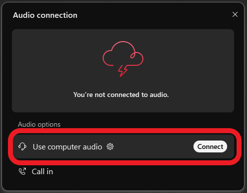
If you have connected by a browser you may need to give your browser access to your microphone in order to connect to the audio.
Click the padlock in the top left of your browser and make sure microphone access is allowed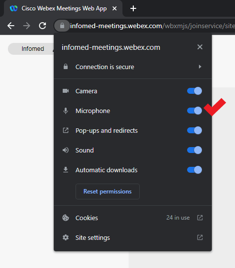
If this does not resolve your issue please email us or call us on 0204 520 5081
What do I need to join a course?
To join an Infomed Online course you simply need an internet connection and a browser (Google Chrome, Mozilla Firefox, Apple Safari).
You can also connect from a mobile device: Download the Webex Meetings app from your App Store.
To join a course with a smooth experience, your internet connection must be stable, not connected to a VPN and at least 20Mbps download.
Below you can use the tool to run an internet speed test.
You must test from:
- — the location that you intend the see the course from;
- — withing the location, if using Wi-Fi, the room or department area that you intend to view the course from to ensure a good signal
- — if connecting from home, a computer that is not connected to a workplace VPN
Speed test
Internet Speed Test
Please test your connection speed at www.fast.com
To join a course with a smooth experience, your internet connection must be stable, not connected to a VPN and at least 20Mbps download.
Fee: £295
- 120 days of access with unlimited playback during this time
- Access to PACS included
- CPD Certificate of attendance upon completion with 12 CPD points
- Opportunity to submit questions directly to the faculty
